Νεύρωμα Morton (Morton’s Neuroma)
Morton’s neuroma is an enlarged nerve that usually occurs in the third interspace, which is between the third and fourth toes (see image below).
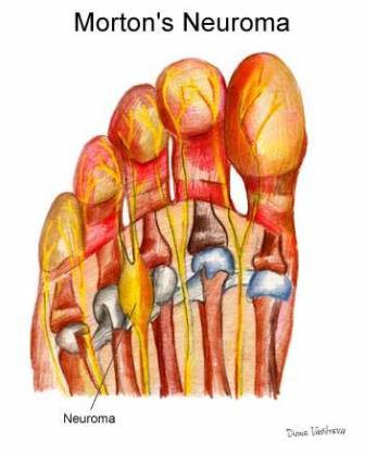
Problems often develop in this area because part of the lateral plantar nerve combines with part of the medial plantar nerve here. When the two nerves combine, they are typically larger in diameter than those going to the other toes. Also, the nerve lies in subcutaneous tissue, just above the fat pad of the foot, close to an artery and vein
Above the nerve is a structure called the deep transverse metatarsal ligament. This ligament is very strong, holds the metatarsal bones together, and creates the ceiling of the nerve compartment. With each step, the ground pushes up on the enlarged nerve and the deep transverse metatarsal ligament pushes down. This causes compression in a confined space.
The reason the nerve enlarges has not been determined. Flatfeet can cause the nerve to be pulled toward the middle (medially) more than normal, which can cause irritation and possibly enlargement of the nerve. The syndrome is more common in women than men, possibly because women wear confining shoes more often. High heels cause more weight to be transferred to the front of the foot and tight toe boxes create lateral compression. As a result, there is more force being applied in the area and the nerve compartment is squeezed on all sides. Under such conditions, even a minimal enlargement in the nerve can elicit pain.
Signs and Symptoms
The most common symptom of Morton’s neuroma is localized pain in the interspace between the third and fourth toes. It can be sharp or dull, and is worsened by wearing shoes and by walking. Pain usually is less severe when the foot is not bearing weight.
Diagnosis
To diagnose Morton’s neuroma the orthopaedic commonly palpates the area to elicit pain, squeezing the toes from the side. Next he or she may try to feel the neuroma by pressing a thumb into the third interspace. The podiatrist then tries to elicit Mulder’s sign, by palpating the affected interspace with one hand and squeezing the entire foot at the same time with the other hand. In many cases of Morton’s neuroma, this causes an audible click, known as Mulder’s sign.
An x-ray should be taken to ensure that there is not a fracture. X-rays also can be used to examine the joints and bone density, ruling out arthritis (particularly rheumatoid arthritis and osteoarthritis).
An MRI scan (magnetic resonance imaging) is used to ensure that the compression is not caused by a tumor in the foot. An MRI also determines the size of the neuroma and whether the syndrome should be treated conservatively or aggressively.
Treatment
In most cases, initial treatment consists of padding and taping to disperse weight away from the neuroma. If the patient has flatfeet, an arch support is incorporated. The patient is instructed to wear shoes with wide toe boxes and avoid shoes with high heels. An injection of local anesthetic to relieve pain and a corticosteroid to reduce inflammation may be administered. The patient is advised to return in a week or 2 to monitor progress. If the pain has been relieved, the neuroma is probably small and caused by the structure of the patient’s foot and the type of shoes the patient wears. It can be relieved by a custom-fitted orthotic that helps maintain the foot in a better position.
Another type of therapy that may be used is alcohol sclerosing injections. In this treatment, the doctor injects a small amount of alcohol in the area of the neuroma area to help harden (sclerose) the nerve and relieve the pain. Injections are given every 7–10 days and, in many cases, 4–7 injections are needed for maximum relief. Please ask your physician for more information regarding this type of treatment.
Conservative treatment does not work for most patients and minor surgery usually is necessary. Two surgical procedures are available.
The dorsal approach involves making an incision on the top of the foot. This approach permits the patient to walk soon after surgery because the stitches are not on the weight-bearing side of the foot. The orthopaedic maneuvers the instruments carefully through many structures and cuts the deep transverse metatarsal ligament, which typically causes most of the nerve compression. This procedure can lead to instability in the forefoot that may require attention in the future.
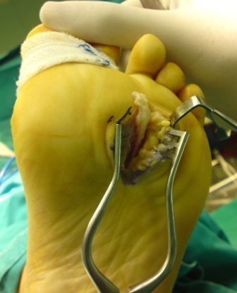
The second procedure involves a plantar approach, in which the incision is made on the sole of the foot. The patient must use crutches for about 3 weeks and the scar that forms can make walking uncomfortable. The advantage of the plantar approach is that the neuroma can be reached easily and resected without cutting any structures.
Surgical Complications
The surgical area contains very small blood vessels, nerves, and muscles and complications can occur. Once the neuroma is removed, the empty space may fill with blood, resulting in a painful hematoma. There is a risk for infection, necessitating careful monitoring by the podiatrist and patient. If the incision site becomes warm or red within a day or two after surgery, or if the patient runs a fever, the surgeon must be contacted immediately.
Recurrence is another possibility. The stump of nerve remaining after resection can begin to grow again. If this occurs, the nerve grows in width and length, creating a burning pain that can be treated by injection or further surgery.